Echocardiogram: diagnosing and monitoring heart conditions through ultrasound
An ultrasound is a specific imaging tool used to diagnose and monitor a wide range of health conditions, including everything from pregnancy to tendon damage to heart disease. A probe that emits ultrasound beams is used to capture real-time images on a screen. Unlike x-rays or CT scans, there is no radiation involved in an ultrasound.
At the Oklahoma Heart Hospital, there are two primary types of ultrasounds of the heart: a transthoracic echocardiogram and a transesophageal echocardiogram.
Transthoracic
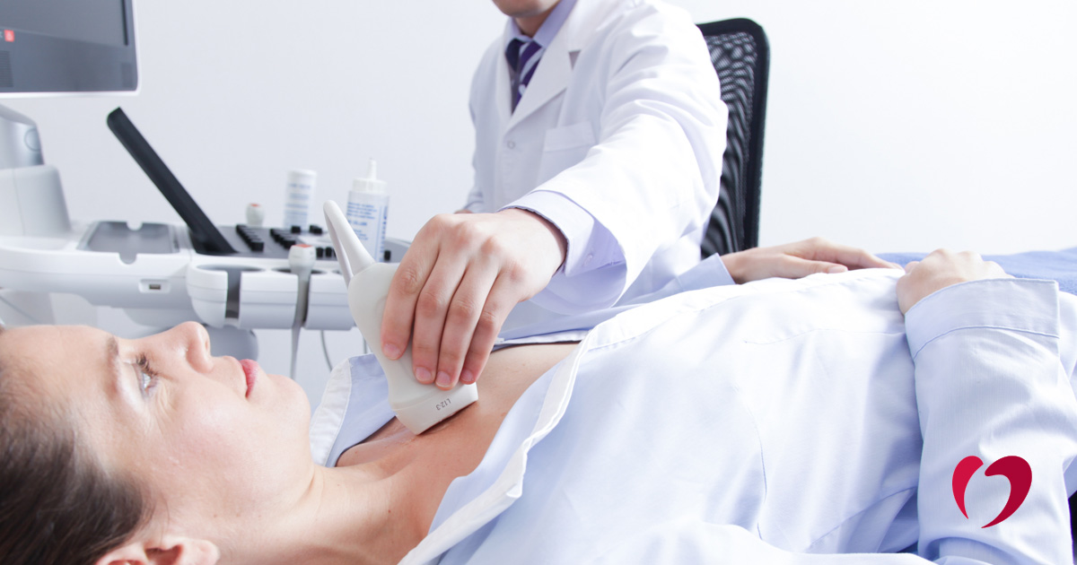
The most commonly used heart ultrasound is the transthoracic echocardiogram (opens in new tab), which is completed with the probe on the outside of the patient’s chest. This imaging test can be used to look at the size and overall function of the heart, assessment the heart valves and pressures in the heart, in addition to the presence of fluid or clots, and more. In other words, a quick and seemingly simple ultrasound of the heart can provide a significant amount of information.
A transthoracic echocardiogram is an extremely common in-office procedure that provides an abundance of information in a short period of time, as it can be used to capture images from four or five different angles of the heart. Your Oklahoma Heart Hospital physician may recommend an echocardiogram due to symptoms or as a follow up to an ECG, or electrocardiogram, which measures the electrical activity of the heart.
Transesophageal
To perform a transesophageal ultrasound (opens in new tab), a probe is placed down the patient’s food pipe to take images from inside the body. Because of the heart’s position near the esophagus, the transesophageal echocardiogram provides us with excellent images of the heart. While much less commonly used than a transthoracic ultrasound, it is especially useful for capturing images of the left side of the heart without any interference from lung tissue or bone.
For electrophysiology concerns, this test can be used to look for clots that may form in the top part of the heart or in the left atrial appendage, which you can’t see as well from a transthoracic ultrasound. The transesophageal ultrasound may also be used as a part of stroke workup, looking for infection of the heart and assessment of valvular heart disease.
A transesophageal ultrasound requires fasting prior to the procedure. The patient will be given an IV for conscious sedation, which will make the patient sleepy. Vital signs are closely monitored during the procedure. Most people do not remember the procedure, but some may intermittently wake up during the study. We use the same type of sedation during transesophageal echocardiograms as one would receive for a colonoscopy.
Depending on what the ultrasound is looking for, it can take anywhere from five to thirty minutes to complete the study. Once the camera is removed, the patient recovers in the hospital for a little while and then goes home. Because the procedure requires numbing the back of the throat, patients should avoid eating for two hours afterward. Some patients may have a sore throat for a day or two following the procedure.
If you are experiencing symptoms or have a family history of heart disease, contact the Oklahoma Heart Hospital today for more information about ultrasound and other diagnostic tools and to schedule an appointment.